
Figure 5.
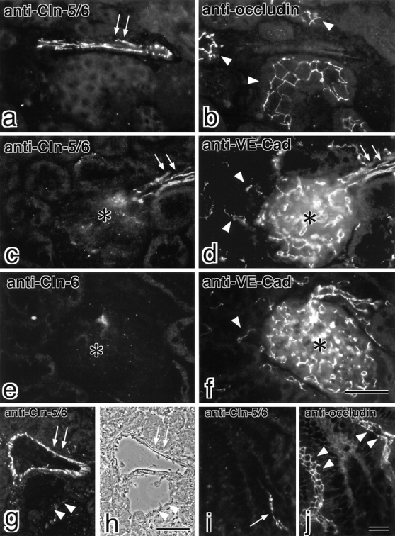
Localization of claudin-5/TMVCF in the kidney and the intestine. Frozen sections of the kidney (a–h) and the intestine (i and j) were double labeled with anti–claudin-5/6 pAb (a) and antioccludin mAb (b), anti–claudin-5/6 pAb (c) and anti–VE-cadherin mAb (d), anti–claudin-6 pAb (e) and anti–VE-cadherin mAb (f), or anti–claudin-5/6 pAb (i) and antioccludin mAb (j). Since anti–claudin-6 pAb yielded no signals in the kidney (e) and the intestine (data not shown), the anti–claudin-5/6 pAb staining can be considered to represent the distribution of claudin-5/TMVCF. Claudin-5/TMVCF appeared to be expressed in a subset of blood vessels (arrows in a and i) that were occludin-negative. Occludin was concentrated at TJs in distal tubules in the kidney (arrowheads in b) and intestinal epithelial cells (arrowheads in j). In the kidney, VE-cadherin–positive intertubular capillaries (arrowheads in d and f) and glomerular capillaries (asterisk in d and f) did not express claudin-5/TMVCF, but afferent and efferent arterioles (arrows in c and d) expressed both claudin-5/TMVCF and VE-cadherin. When transverse frozen sections of the thicker artery and vein (arrows and arrowheads, respectively, in g and h) of the kidney were stained with anti–claudin-5/6 pAb, only the artery was intensely stained (g). (h) Phase–contrast image. Bars: 40 μm for a–f (f); 70 μm in g and h (h); 40 μm in i and j (j). Cln, claudin; Cad, cadherin.