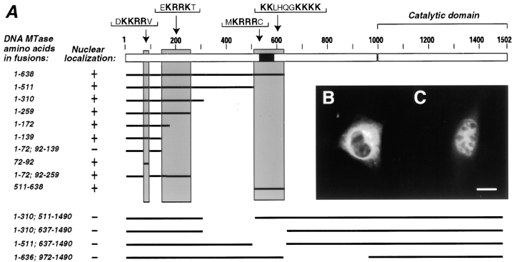
Figure 2.
Dnmt1 has at least three independent nuclear localization signals. (A) Diagram outlining the structure of the oocyte-specific murine Dnmt1 protein and indicating the different parts of the Dnmt1 present in fusions with the β-galactosidase epitope (for simplicity, the latter is not depicted). The different fusion constructs were expressed in murine myoblast or fibroblast cells, formalin fixed, stained with anti–β-galactosidase monoclonal antibody, and screened for their nuclear or cytoplasmic localization. B shows a representative example of a cytoplasmic fusion protein containing amino acids 1-72; 92-139 of Dnmt1 and C shows a nuclear fusion protein containing amino acids 1-72; 92-259 of Dnmt1, including only the second NLS. The analysis of all listed constructs lead to the identification of three regions (highlighted by grey shading) that contain independently functional NLS. Sequences representing possible NLS within each shaded area are specified below. The black box indicates the location of the cysteine-rich region that had been shown to bind zinc ions (Bestor 1992). Bar, 10 μm.

