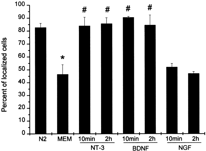
Figure 2.
Quantitative analysis of neurotrophin stimulated β-actin mRNA localization. Neurotrophin-3 (NT-3), brain-derived neurotrophic factor (BDNF), or NGF was added to the MEM (25 ng/ml) for the indicated time. NT-3 and BDNF, but not NGF, were observed to rapidly stimulate the localization of β-actin mRNA within growth cones. The axon-like neurite and growth cone from each cell was visualized for the presence of β-actin mRNA granules (see Materials and Methods). 100 cells were scored per coverslip. Histogram shows mean percentage and standard deviation between independent samples (n = 4). Examples of localized and nonlocalized cells are shown in Fig. 1. N2, normal control. MEM, starvation in minimum essential medium. *, P < 0.01 when MEM compared with N2. #, P < 0.01 when NT-3 or BDNF was compared with MEM.

