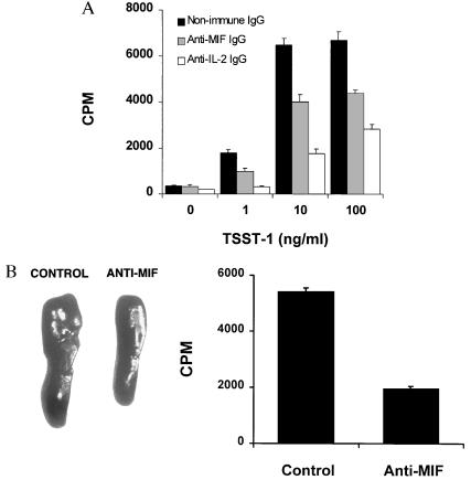
Figure 4.
Anti-MIF antibody inhibits TSST-1-induced proliferation of mouse splenocytes. (A) Effect of anti-MIF antibody on the proliferation of splenocytes stimulated by TSST-1 in vitro. Splenocytes (4 × 10⁵ cells) from BALB/c mice were stimulated for 72 hr with TSST-1 at the indicated concentrations in the presence of neutralizing anti-MIF IgG (50 μg/ml)(stippled columns), nonimmune IgG (50 μg/ml) (solid columns), or anti-IL-2 IgG (5 μg/ml) (open columns). Cells then were pulsed with [3H]thymidine (0.5 μCi/well) for 6 hr and the incorporation of thymidine measured by liquid scintillation counting. Data from one representative experiment are shown and expressed as the mean ± SEM of quintuplicate values. (B). Effect of anti-MIF antibody on the proliferation of splenocytes stimulated by TSST-1 in vivo. C57BL/6 mice were injected i.p. with 200 μl of anti-MIF or nonimmune (control) rabbit serum 2 hr before 20 μg of TSST-1. After 3 days, spleens were harvested and weighed. Spleens of two representative mice treated with nonimmune serum or anti-MIF serum are shown in the left panel. Splenocytes (5 × 10⁵ cells per well) were prepared as described in Materials and Methods, pulsed for 6 hr with [3H]thymidine (0.5 μCi/well) and the incorporation of thymidine measured by liquid scintillation counting. Data from three separate experiments (n = 8 mice per treatment group) are expressed as the mean cpm ± SD of splenocyte cultures tested in quintuplicate (Right).

