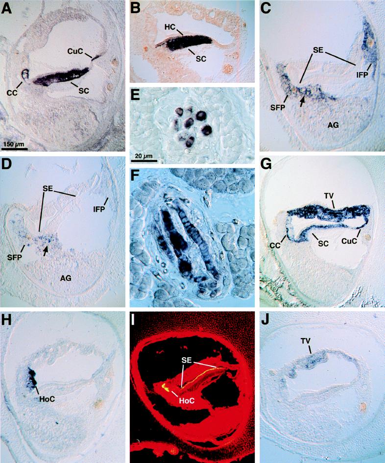
Figure 1.
In situ hybridization analysis of cDNAs expressed in the inner ear. (A) On a cross-section of the basilar papilla, β-tectorin mRNA occurs in clear cells (CC), supporting cells (SC), and cuboidal cells (CuC). (B) Calbindin mRNA is abundant in supporting cells (SC) and detectable in hair cells (HC). (C) Strong signals for type II collagen mRNA are visible in the superior fibrocartilaginous plate (SFP) and inferior fibrocartilaginous plate (IFP) and, as indicated by the arrow, between the auditory ganglion (AG) and sensory epithelium (SE). All collagen mRNAs found in the inner ear were expressed in a similar manner. (D) Coch-5B2 mRNA occurs in spindle-shaped cells (arrow) marking the path of neurites between the auditory ganglion (AG) and sensory epithelium (SE). Weaker expression is detectable in the superior and inferior fibrocartilaginous plates (SFP and IFP). (E and F) Coch-5B2 mRNA displays strong labeling of muscle spindles in the gastrocnemius muscle; note the encapsulation and the nuclear chain in (F). (G) Connexin 31 mRNA is expressed robustly in cells of the tegmentum vasculosum (TV), cuboidal cells (CuC), supporting cells (SC), and clear cells (CC). (H) Homogenin mRNA is detectable in homogene cells (HoC). (I) The intense yellow fluorescence of rhodamine-coupled phalloidin signals a high abundance of filamentous actin in homogene cells (HoC) as well as in hair bundles of the sensory epithelium (SE). (J) Otokeratin mRNA occurs in cells of the tegmentum vasculosum (TV).

