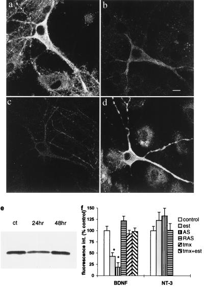
Figure 1.
BDNF immunoreactivity is down-regulated by estradiol in cultured hippocampal neurons. (a) Control. (b) Estradiol, 24 hr. AS-treated (c) cells exhibit a marked reduction in staining for BDNF whereas RAS-treated cells (d) display normal staining. (Bar = 10 μm.) (e) Typical western blot of control cultures (left) and those collected after 24- and 48-hr exposure to estradiol. Lanes were loaded with equal protein, and a single BDNF band appeared near 14 kDa. On average (3 experiments), the density of the BDNF band at 24 hr was 64% of control. (f) BDNF fluorescence staining intensity measurements from several experiments were averaged and presented as a percent of control, which was run separately for each experiment. Bars represent means ± SEM, and asterisks represent statistically significant difference from control at P < 0.01, Student’s t test. The bars, from left to right, are control, estradiol (est), BDNF-AS, BDNF-RAS, tamoxifen (tmx), and tamoxifen + estradiol (tmx + est). Only the estradiol and the BDNF-AS caused a significant reduction in BDNF staining. On the right are measurements of neurotrophin-3 (NT-3) fluorescence intensity, showing little effect of estradiol or the BDNF-AS.

