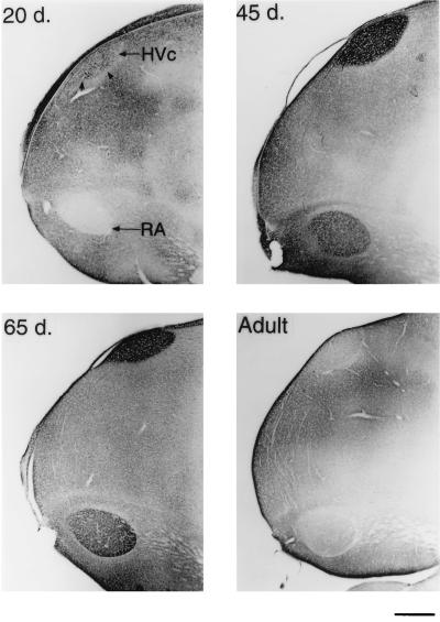
Figure 2.
Sagittal sections of the caudal telencephalon stained for BDNF. A 20-day-old male has many BDNF containing cells, but little staining is seen in RA (Upper Left). By 45 days, BDNF staining is pronounced in HVc cells and neuropil, some of the axon bundles that connect HVc and RA, and in the RA neuropil only (Upper Right). In a 65-day-old male, BDNF staining within HVc is still heavy, but is mostly confined to the neuropil. The RA is also heavily labeled at this age (Lower Left). In the adult, however, BDNF immunostaining within these song nuclei is sharply reduced (Lower Right). (Bar = 500 μm.)

