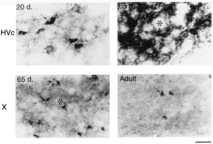
Figure 5.
The different immunostaining patterns within HVc and area X of the developing male zebra finch. At 20 days of age, HVc contains many BDNF stained neurons and fine processes (Upper Left). At 65 days, however, only a few HVc cell bodies are labeled, but most of the staining shifts to the neuropil and/or extracellular matrix surrounding the unstained cells (asterisk, Upper Right). Also at 65 days, BDNF labeling in area X is rapidly increased and appears clustered around unlabeled cell bodies (asterisk, Lower Left). In the adult, however, there are many widely spaced neurons in area X that contain BDNF surrounded by many small knob-like objects (Lower Right). (Bar = 20 μm.)

