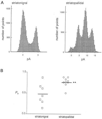
Figure 3.
Comparison of fractional open probabilities of the D2 dopamine receptor-activated K+ channel on identified striatonigral and striatopallidal neurons. (A) Examples of all-points amplitude histograms of a striatonigral cell with one active channel in the patch (Left) and a striatopallidal cell with three active channels in the patch (Right). In each case, the relative amplitude of the peak at 0 pA is proportional to the fraction of time spent in the closed state. (B) Distribution of fractional open probability (Po) values for striatonigral and striatopallidal neurons. Each point represents the value for a different cell at resting membrane potential. Horizontal lines are the means of these values; ∗∗, value significantly greater than for striatonigral neurons (P < 0.01).

