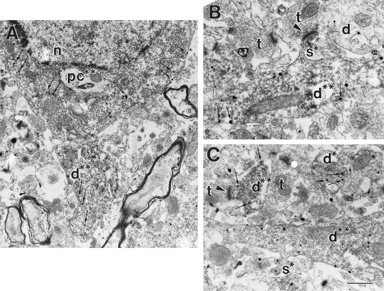
Figure 5.
The m1-muscarinic receptor colocalizes with the NMDA NR1 subunit. Double-labeling electron microscopic immunocytochemistry was used to identify immunoreactivity for the m1 mAChR (visualized with silver-enhanced immunogold, electron-dense round particles) and the NR1 subunit of the NMDAR protein (visualized with DAB; a diffuse floccular reaction product). (A) A pyramidal cell soma (pc**) that contains immunoreactivity for m1 and NR1. Note the presence of DAB (dark diffuse-reaction product filling cytoplasm, and immunogold particles present in the cytoplasm and lining the membrane highlighted with small arrows in A–C). Also pictured is a large proximal dendrite (d**) double-labeled with both m1 and NMDAR immunoreactivity. n, nucleus. (B) A large proximal dendrite (d**) that contains immunoreactivity for both the m1 and NMDARs. A dendritic spine (s*) containing NMDA immunoreactivity is seen extending off the proximal dendrite that is receiving an asymmetric (excitatory) synapse (arrowhead) from an unlabeled synaptic terminal (t). An unlabeled dendrite (d) is also pictured. (C) Several proximal and distal dendrites that are double-labeled for m1 and NMDAR proteins (d**) are shown. The two dendrites at the top of the panel are smaller distal dendrites, while the one at the bottom is a larger proximal dendrite. The distal dendrite on the top left can be seen receiving an asymmetric synapse (arrowhead) from an unlabeled terminal (t). Additionally, a dendritic spine (s*) containing immunoreactivity for the m1 receptor is shown extending off of the proximal dendrite. [Bar = 1.2 μm (A); 465 nm (B); 440 nm (C).]

