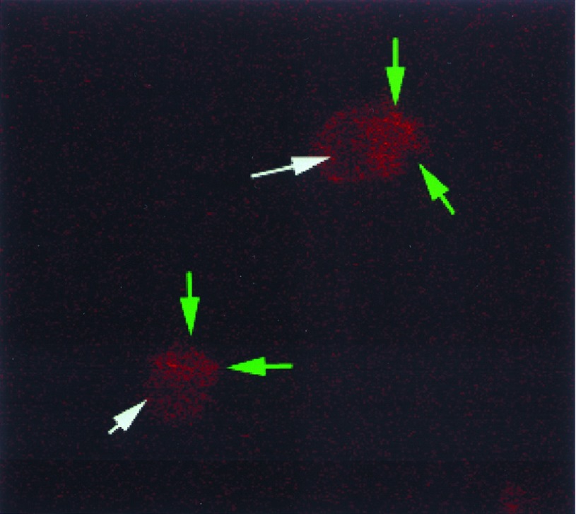
Figure 5.
Confocal image of K562 cells injected with AS vav MBs. Cells injected with AS vav MBs revealed fluorescence signal when irradiated by a laser tuned to excite at 351 nm. Fluorescence images were gathered 15–30 min after MB injection and appeared stronger in the cells’ cytoplasm (outlined by green arrows) than in the nucleus (white arrow), suggesting that hybridization may be favored in the latter location. Uninjected cells or cells injected with control MBs displayed little or no signal and were, therefore, very dark or unseen.

