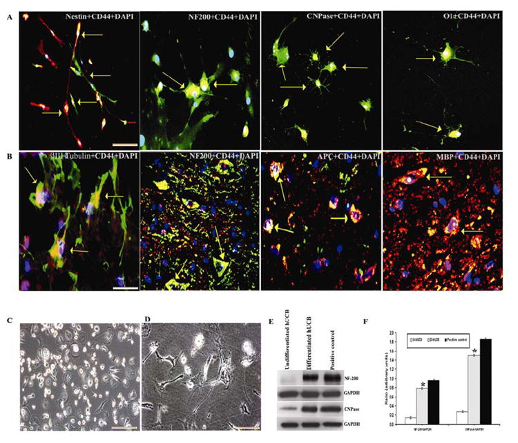
Fig. 1. Trans-differentiation of hUCB into neural phenotypes.

(A) Photomicrographs of hUCB in mixed culture demonstrating neural proteins expressed by the cells before transplantation. FITC-conjugated- Nestin and NF-200 (for neurons) with Texas-red conjugated- CD44 (for hUCB); Texas-red conjugated –CNPase and O1 (for oligodendrocytes) with FITC-conjugated- CD44 (for hUCB). Cells displaying neuron like morphology with long axonal projections, CNPase-immunoreactive cells displaying morphology characteristic of oligodendrocytes, with flat cell body and short or long branched projections. (B) Confocal immunohistochemistry on longitudinal sections of the injured spinal cords of rats 5 weeks after transplantation is shown. Immunofluorescence analysis of cryo-sections indicates co-localization (yellow) of FITC-conjugated –βIII tubulin and NF-200 (for neurons) with Texas-red conjugated- CD44 (for hUCB); FITC conjugated-CD44 (for hUCB) with Texas-red conjugated -APC and -MBP (for oligodendrocytes) as shown by (↑). The results are from 3 independent sections between 1 and 2mm from the injury epicenter from treated rats (n =5). Bar = 100 μm. (C) Phase-contrast image of undifferentiated hUCB. (D) Phase-contrast image of differentiating hUCB after 4 days showing mixed population of differentiated neural phenotypes. (E) Western blot showing neural proteins in differentiated population. Equal amounts of protein (40 μg) were loaded onto 10%-14% gels and transferred onto nylon membranes, which were then probed with respective antibodies. The blots were stripped and reprobed with GAPDH to assess protein levels. (F) Quantitative estimation of neural proteins in Fig. F. (U-hUCB = Undifferentiated hUCB; D- hUCB = Differentiated hUCB; Positive control = Whole brain extract of rat). Results are from 3 parallel experiments from 3 separate cord blood preparations. A subpopulation of hUCB-derived cells growing in a monolayer before differentiation was found to be negative for all investigated antigens. Merged figures include colocalized markers, shown by (↑). For panels A, B and C, Bar = 100 μm. For panel D, Bar = 200 μm. (Error bars indicate SEM. * Significant at p <0.05).
