
Fig. 3. Fas immunoreactivity on TUNEL positive cells.
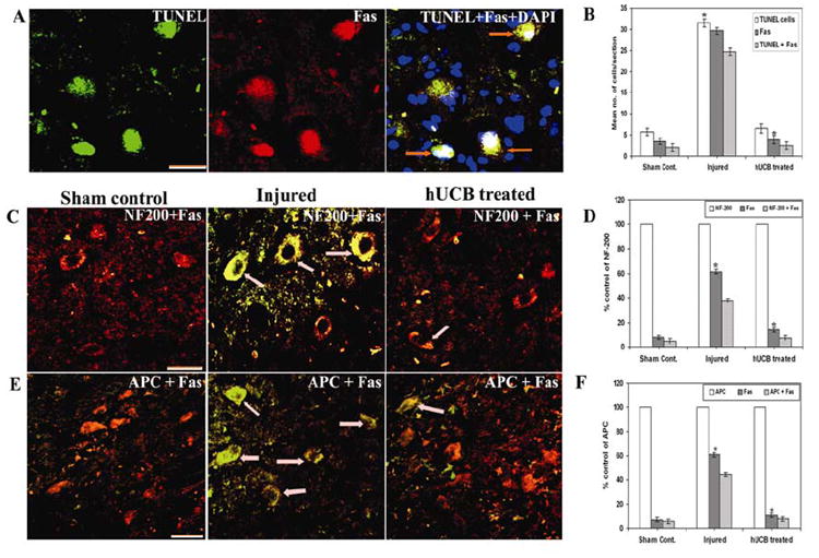
(A) Expression of Fas (Texas-red conjugated) on TUNEL positive cells (green) from injured sections. (B) Quantitative estimation of TUNEL, Fas and TUNEL + Fas cells in injured and treated groups. (C) Cryo-sections showing Co-localization of Fas and NF-200 (specific for neurons) and (E) Fas and APC (mature marker for oligodendrocytes) established the expression of Fas on neurons and oligodendrocytes (↑) undergoing apoptosis. Fas is FITC-conjugated and NF-200 and APC are Texas-red conjugated. Note a significant decrease in the expression of Fas in hUCB-treated sections. For panels A, C and E Bar = 100 μm. (D) Quantification of NF-200 + Fas cells and (F) APC + Fas cells. (Error bars indicate SEM. * Significant at p <0.05). A total of more than 100 cells were counted per each field and more than five fields were chosen at random on each section. Results are from three sections between 1 and 2 mm caudal to the injury epicenter after 3 weeks SCI (n ≥ 3).