
Fig. 4. Immunoblot analysis of Fas and other death ligands in spinal cord sections.
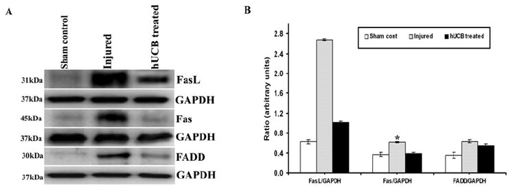
Equal amounts of protein (40 μg) were loaded onto 10%-14% gels and transferred onto nylon membranes, which were then probed with respective antibodies. The blots were stripped and reprobed with GAPDH to assess protein levels. A shows the FasL, Fas and FADD proteins with respect to GAPDH; B shows their quantitative estimations. Inhibition of the apoptotic pathway by hUCB shows downregulation of FasL, Fas and FADD. Each blot is representative of experiments performed in duplicate with each sample (n ≥ 3). Error bars indicate SEM. * Significant at p <0.05.