
Fig. 7. Immunoblot analysis of activated inhibitors of apoptosis in spinal cord sections.
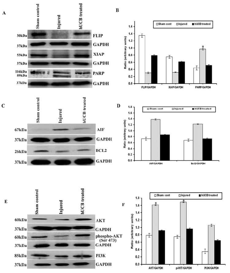
Equal amounts of protein (40 μg) were loaded onto 10%-14% gels and transferred onto nylon membranes, which were then probed with respective antibodies. The blots were stripped and reprobed with GAPDH to assess protein levels. A and C shows the inhibitors of apoptosis present in cytosol and nucleus whereas E shows the survival signaling pathway proteins with respect to GAPDH; B, D and F are their quantitative estimations, respectively. Upregulation of inhibitory proteins FLIP, XIAP, and inhibition of PARP cleavage is clearly seen hUCB treatments. Each blot is representative of experiments performed in duplicate with each sample (n ≥ 3). Error bars indicate SEM. * Significant at p <0.05.