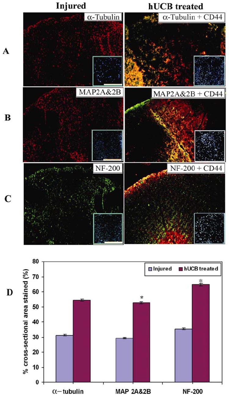
Fig. 8. Structural integrity of the injured spinal cord maintained by hUCB after intraspinal grafting.

Immunofluorescence studies of spinal cord cryo-sections showing dorsal white matter with cytoskeletal markers viz., α-tubulin (A), MAP2A & 2B (B) and NF200 (C) were performed. α-tubulin, MAP2A & 2B are Texas-red conjugated and their respective CD44 are FITC-conjugated. NF-200 is FITC-conjugated and CD44 are Texas-red conjugated. Inset shows corresponding DAPI images. Bar = 2000μm. (D) Quantitative estimation of the cross-sectional area stained. Calculation of % staining was done on only the lineage specific markers, (but not on the merged image) so that exact comparison with left panel images can be done. When compared to the injured sections, hUCB-treated sections have a high level of structural integrity of the spinal cord. Values represent mean ± SEM values of at least three sections with duplicates from each condition. * Significant at p <0.05. Results are from 3 independent sections between 1 and 2mm from the injury epicenter after 3 weeks SCI (n ≥ 3).
