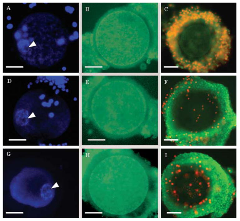
Fig. 1.

Structural integrity of grade I immature oocyte during or after exposure to hypo- (100 mOsm; A–C), iso- (290 mOsm; D–F), and hyperosmotic (2,000 mOsm; G–I) solutions. A,D,G: Germinal vesicle chromatin stained with Hoechst 33341 containing nucleolus (white arrow) during exposure. B,E,H: Mitochondrial distribution assessed with Mitotracker green after exposure. C,F,I: Membrane integrity of cumulus cells (intact cells stained with green SYBR14, non-intact cells stained with propidium iodide) after exposure. Bar = 50 μm. [See color version online at www.interscience.wiley.com.]
