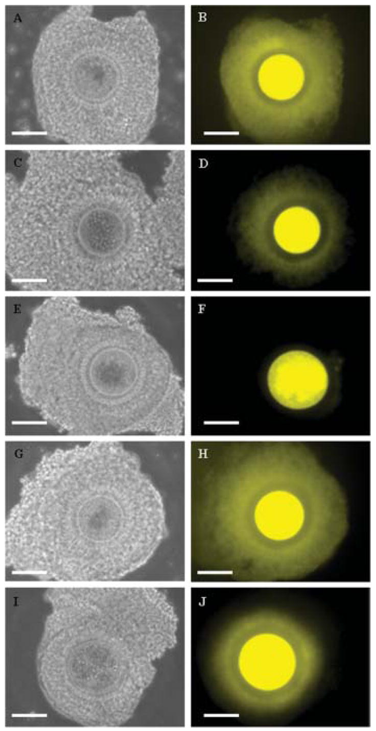
Fig. 2.

Structural integrity of grade I immature oocytes (bright field microscopy; A,C,E,G,I) and cumulus–oocyte communications (micro-injection and diffusion of fluorescent Luciferin Yellow; B,D,F,H,J) after exposure to anisosmotic and isosmotic solutions. A,B: Cumulus–oocyte complex (COC) with open communications after exposure to hypo- or isosmotic condition. C,D: COC with partially open communications after exposure to 1,000 mOsm. E,F: COC with closed communications after exposure to 2,000 mOsm. G,H: COC with open communications after treatment with cytochalasin B and exposure to 1,000 mOsm. I,J: COC with partially open communications after treatment with cytochalasin B and exposure to 2,000 mOsm. Bar = 100 μm. [See color version online at www.interscience.wiley.com.]
