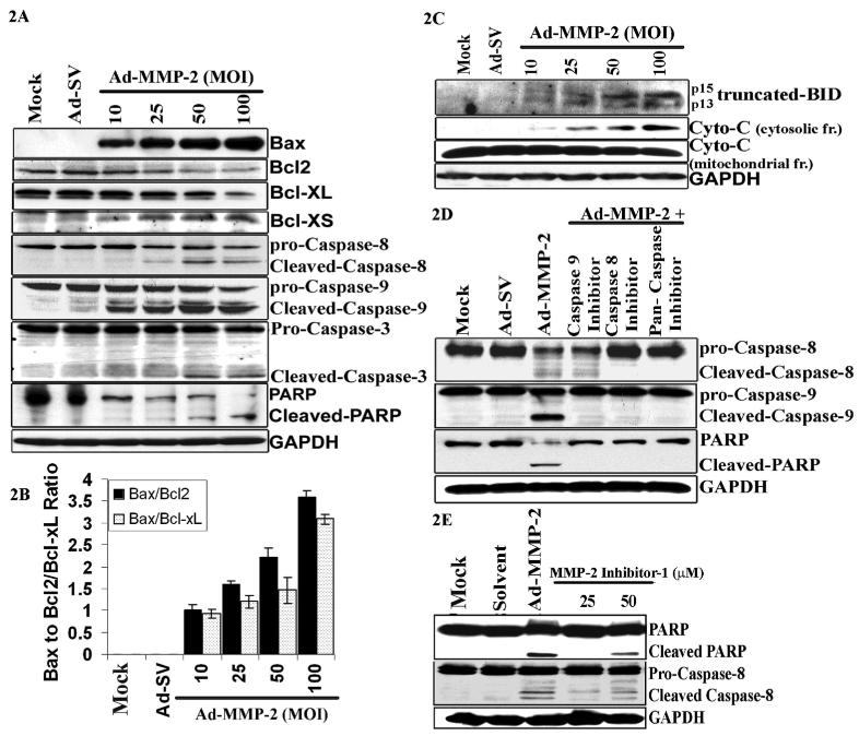
Figure 2. Ad-MMP-2 infection induces cleavage of caspases and PARP.
A549 cells were infected with mock, 100MOI of Ad-SV or the indicated dose of Ad-MMP-2 for 48h. (A) Cell lysates were used for immuno blot analysis of Bcl-2 family proteins and caspases. (B) Densitometric analysis showing the Bax/Bcl2 and Bax/ Bcl-xL ratios in treatment cells. The, bars indicate standard errors (±SE) from the mean of three separate experiments. (C) Cells were used to evaluate Bid cleavage and cytochrome-c release in the cytosol. (D) The effects of caspase inhibitors on Ad-MMP-2-induced apoptosis. A549 cells were treated with 100μM of various caspase inhibitors or solvent (DMSO) for 1 h and infected with mock, 100MOI of Ad-MMP-2. Caspase cleavage and PARP levels were determined by immuno blotting. Anti-GAPDH antibody was used for visualizing protein loading. immuno blots are representative of three experiments. (E) Cleavage of caspase-8 and PARP-1 in A549 cells treated with MMP-2 inhibitor-1, a MMP-2 specific inhibitor (50μM), indicating that MMP-2 inhibition induces apoptosis.

