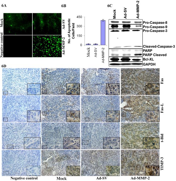
Figure 6. Ad-MMP-2 treatment induces apoptosis in A549 subcutaneous tumors.
(A) Tumor sections were stained for apoptosis by TUNEL staining. Data shown are representative fields. (B) Quantitation of apoptotic cells indicated an increase in number of TUNEL-positive cells per microscopic field in Ad-MMP-2 treated cells. Bars represent the mean and standard error of the mean of three sections from three animals of the same treatment. (C) Processing of PARP and caspases-8, -9 and -3 was detected in total cell lysates from the subcutaneous tumors of mice, which received mock, Ad-SV and Ad-MMP-2. Representative results obtained from the tumor of one mouse per treatment condition are shown. (D) Immunohistochemical analysis of Fas, Fas-L, TIMP-3 and Bid showing increased expression of these proteins in Ad-MMP-2-treated tumors compared to mock and Ad-SV controls.

