
Figure 2.
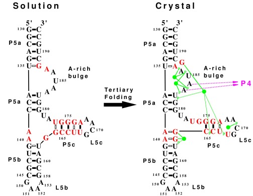
The secondary structures determined by NMR in solution and by x-ray diffraction in a crystal are compared. Nucleotides that change their base pairing are in red. The solution structure represents the unfolded state, whereas the crystal structure represents the folded state with tertiary interactions. Each solid bar denotes a Watson–Crick base pair, and each hollow bar denotes a non-Watson–Crick base pair. The tertiary interactions seen in the crystal (10) include binding of A183 and A184 to the minor groove of P4 (shown as purple arrows). The green disks represent five magnesium ions identified in the crystal (9, 10) that form direct hydrogen bonds (thick green line) and water-mediated hydrogen bonds (thin green lines) with phosphate oxygens (indicated by p) and guanine bases. The tertiary folding causes significant secondary structure rearrangements, including the loss of three G⋅U base pairs, the loss of a GNRA tetraloop, the formation of a single base bulge, and the addition of two G⋅A base pairs.