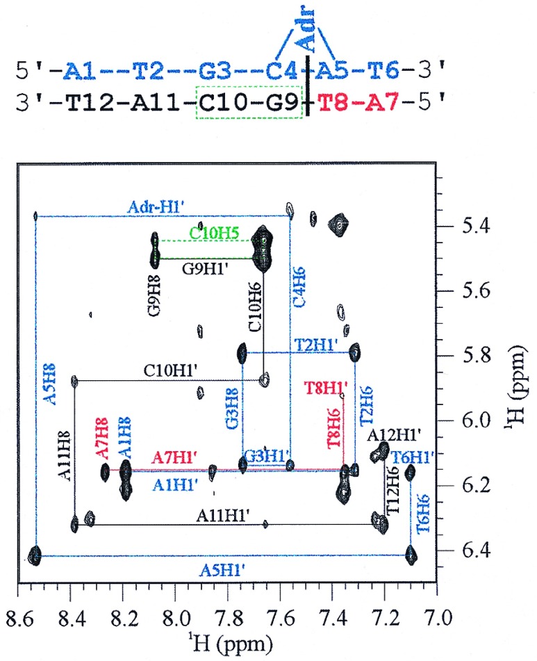
Figure 3.
Anomeric-aromatic portion of the 500-Mhz 1H NOESY spectrum at 10°C of the Adriamycin-DNA adduct mediated by Tris/Fe(III)/DTT. The connectivity patterns between anomeric and aromatic DNA protons are correlated by color to the schematic Adriamycin-DNA adduct shown above. The c-strand is shown in blue, whereas the n-strand is depicted in black and red. The NOESY connectivity pattern between DNA protons is interrupted between the C4pA5 step of the c-strand and the T8pG9 step of the n-strand, indicating intercalation of the Adriamycin chromophore at this point and covalent attachment to G3. Connectivity along the c-strand is completed by contacts between the aromatic protons C4H6 and A5H8 to the Adriamycin-H1′ carbohydrate proton. The box representing connectivity between the protons G9H8, G9H1′, C10H6, and C10H5, shown in green, was a characteristic feature of all observed Adriamycin-DNA spectra of the type d[(AT)nGC(AT)n]2. Residual unlabeled cross-peaks represent small amounts of unreacted DNA that was coexcised from the gel with the adduct.

