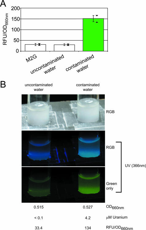
FIG. 4.
PurcA gfpuv reporter detection of uranium-contaminated groundwater. (A) Strain NJH371 GFPuv fluorescence was assayed after 4 h of exposure to uranium-contaminated (4.2 μM uranium) or uncontaminated (<0.1 μM uranium) Oak Ridge Field Research Center water samples. M2G minimal medium was used as a negative control. For an explanation of the error bars and triangles, see the legend to Fig. 3D. The data are aggregate results from four experiments. (B) Cultures of strain NJH371, illuminated with either daylight or a hand-held UV lamp, were photographed after 4 h of exposure to uranium-contaminated or uncontaminated water samples. The isolated green channel of the UV-illuminated RGB image (red, green, and blue channels), as well as culture OD660 and GFPuv fluorescence values for the cultures photographed, are shown. RFU, relative fluorescence units.

