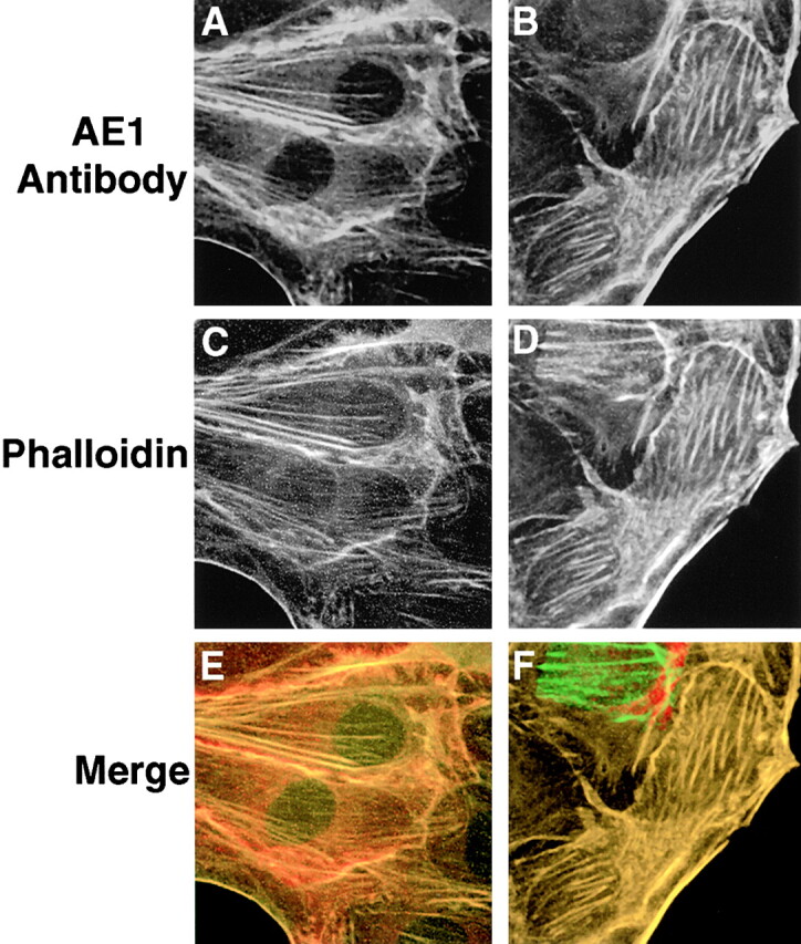
Figure 6.

AE1-4 colocalizes with phalloidin-stained microfilaments in transfected MDCK cells. MDCK cells stably expressing the AE1-4 anion exchanger were grown in subconfluent monolayers. Cells were either untreated (A, C, and E) or extracted with 0.5% Triton X-100 in PBS (B, D, and F) before fixation in 1% paraformaldehyde in PBS. The cells were then incubated with an AE1-specific peptide antibody, followed by incubation with DAR-IgG conjugated to lissamine and FITC-conjugated phalloidin. Images showing the distribution of AE1-4 (A and B) and phalloidin-stained microfilaments (C and D) were collected on a Zeiss Axiophot microscope. The merged images (E and F) were generated in Adobe Photoshop.
