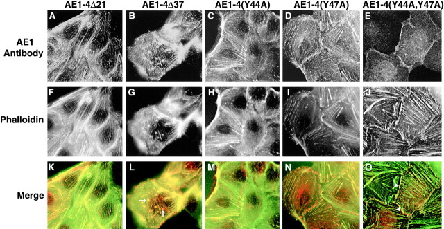
Figure 9.
Localization of AE1-4 mutants in subconfluent MDCK monolayers. MDCK cells expressing AE1-4Δ21 (A), AE1-4Δ37 (B), AE1-4(Y44A) (C), AE1-4(Y47A) (D), or AE1-4(Y44A,47A) (E) were grown on coverslips in subconfluent monolayers. The cells were fixed and incubated with an AE1-specific peptide antibody, followed by incubation with DAR-IgG conjugated to lissamine (A–E) and FITC-conjugated phalloidin (F–J). Immunoreactive polypeptides and phalloidin stained microfilaments were visualized on a Zeiss Axiophot microscope. Images were collected at the basal surface of the cells. Merged images were generated in Adobe Photoshop (K–O). The arrows in L mark two of the actin-containing clusters that colocalize with AE1-4Δ37. The arrowheads in O indicate where AE1-4(Y44A,47A) colocalizes with cortical actin at sites of cell-cell contact.

