
Figure 10.
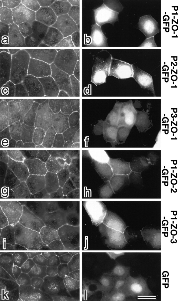
Behavior of GFP-fusion proteins with PDZ1, -2, and -3 domains of ZO-1/ZO-2/ZO-3 in cultured epithelial cells (MDCK cells). GFP (GFP) or GFP-fusion proteins with PDZ1 domain of ZO-1 (P1-ZO-1-GFP), PDZ2 domain of ZO-1 (P2-ZO-1-GFP), PDZ3 domain of ZO-1 (P3-ZO-1-GFP), PDZ1 domain of ZO-2 (P1-ZO-2-GFP) or PDZ1 domain of ZO-3 (P1-ZO-3-GFP) were exogenously and transiently expressed in MDCK cells. These cells were fixed and stained with anti–claudin-1 pAb (a, c, e, g, i, and k) in red. Expressed GFP or GFP-fusion proteins were visualized by green fluorescence (b, d, f, h, j, and l). PDZ1 domains of ZO-1 (b, P1-ZO-1-GFP), ZO-2 (h, P1-ZO-2-GFP), ZO-3 (j, P1-ZO-3-GFP), and PDZ2 domain of ZO-1 (d, P2-ZO-1-GFP) were recruited to claudin-1–positive TJs. PDZ3 domain of ZO-1 (f, P3-ZO-1-GFP) also appeared to be concentrated at TJs, although very faintly as compared with b, d, h, and j. No concentration of GFP at cell–cell borders was observed (l). Bar, 10 μm.