
Figure 7.
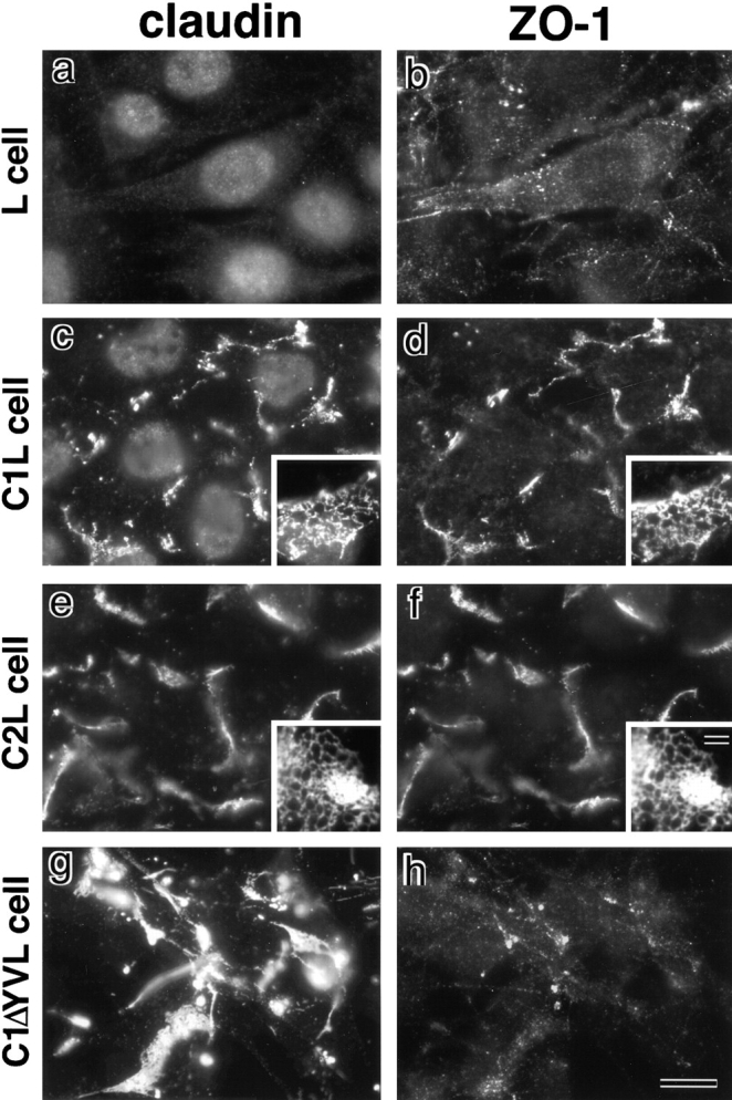
Recruitment of endogenous ZO-1 to claudin-based networks in L transfectants expressing claudin-1 or -2. Parental L cells (L cell; a and b) and L transfectants expressing claudin-1 (C1L cell; c and d) were double stained with rat anti–claudin-1 mAb (claudin; a and c) and mouse anti–ZO-1 mAb (ZO-1; b and d). L transfectants expressing claudin-2 (C2L cell; e and f) were double stained with anti–claudin-2 mAb (e) and anti–ZO-1 mAb (f). L transfectants expressing claudin-1 mutant lacking the COOH-terminal YV sequence (C1ΔYVL cell; g and h) were double stained with anti–claudin-1 pAb (g) and anti–ZO-1 mAb (h). ZO-1 showed no characteristic concentration in L cells (b), whereas in C1L and C2L cells ZO-1 was coconcentrated with claudin-1 (c and d) and claudin-2 (e and f) as planes at cell–cell borders. Close inspection revealed that in these cells both claudins and ZO-1 were concentrated in elaborate network patterns, and that their network patterns were mostly overlapped (insets in c–f). In C1ΔYVL cells, mutant claudin-1 was concentrated at cell–cell borders (g), whereas ZO-1 showed no concentration (h). Bar: (a–h) 10 μm; (insets) 15 μm.