
Figure 3.
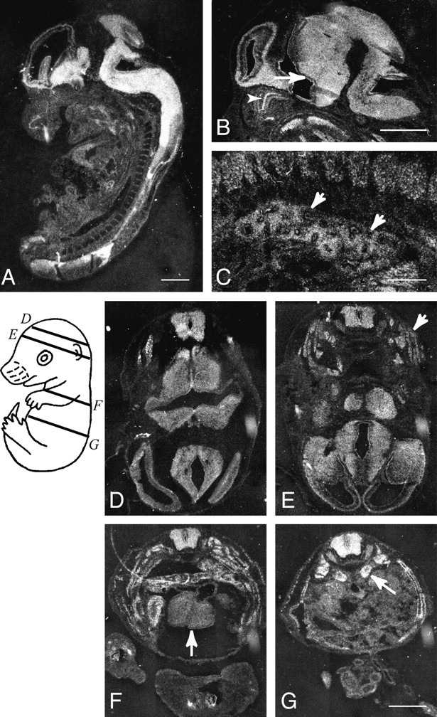
Expression of ACF7 mRNAs in the mouse embryo. Sagittal sections (A–C) and transverse sections (D–G) of an embryonic day 14.5 mouse embryo were hybridized with mACF7 antisense probe. Relative planes of sectioning for sections shown in D–G are illustrated on a schematic drawing of the embryo. In addition to ubiquitous expression of mACF7 mRNA throughout the embryo, high levels of expression are observed in the brain and spinal cord. In the brain, both ependymal layer (B, arrow) and mantle layer are heavily labeled. In the peripheral tissue, an intermediate to high level of ACF7 expression is observed in the dorsal root ganglia, olfactory epithelium (B, arrowhead), intrinsic muscle of the tongue, bronchial epithelium, and mesenchyme of the lung (B, arrows), skeletal muscle of the trunk (E, arrow), myocardium (F, arrow), and adrenal glands (G, arrow). Bars: 1 mm (A, B, D–G); and 0.1 mm (C).