
Figure 5.
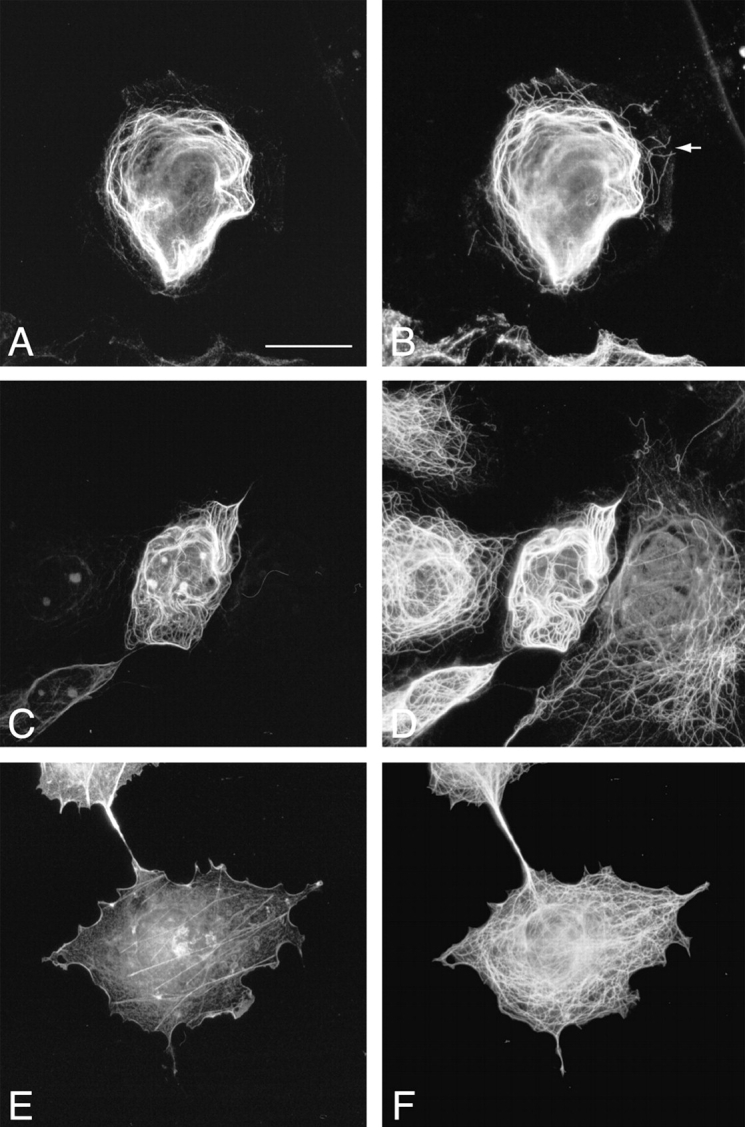
Association of ACF7 COOH-terminal domain with MTs. Transient transfections of pFLAG-mACF7-C (A–D) and pFLAG-ABD (E and F) were performed in COS-7 cells. Transfected cells were stained with monoclonal anti-FLAG M2 antibody (A, C, and E), polyclonal anti–Tyr-tubulin antibody (B), polyclonal anti–Glu-tubulin antibody (D), and polyclonal antitubulin antibody (F), and were examined by immunofluorescence microscopy. mACF7-C proteins colocalize with most of the Tyr-MTs (A and B) and all of the Glu-MTs (C and D). The noncolocalized Tyr-MTs are indicated by an arrow (B). Frequently, long MTs forming whorls were observed in pFLAG–mACF7-C transfected cells (B and D). The overexpressed ABD protein did not associate with the MT network (E and F). Bar, 20 μm.