
Figure 7.
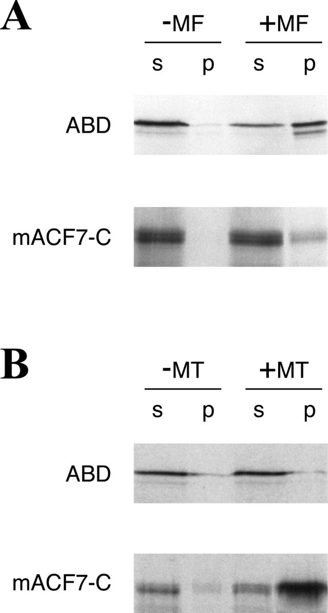
Interaction of mACF7 with polymerized actin and MTs in vitro. (A) Actin spin-down binding assays were performed with [35S]methionine ABD and mACF7-C proteins. In vitro translated mACF7-C protein appeared as a doublet in SDS-PAGE. These two different sized proteins might be the result of degradation or alternative initiation. In the presence of polymerized actin filaments (+MF), a significant portion of ABD was found in the pellet (p). Without actin (−MF), most of the ABD remained in the supernatant (s). A small portion of mACF7-C protein was also found in the pellet, indicating weak interaction between mACF7-C protein and actin filaments. (B) Microtubule spin-down binding assays were performed with [35S]methionine-labeled ABD and mACF7-C proteins. In the presence of polymerized MTs (+MT), mACF7-C but not ABD protein cosediment with the MT pellet (p). Without MTs (−MT), both of the proteins remained in the supernatant (s).