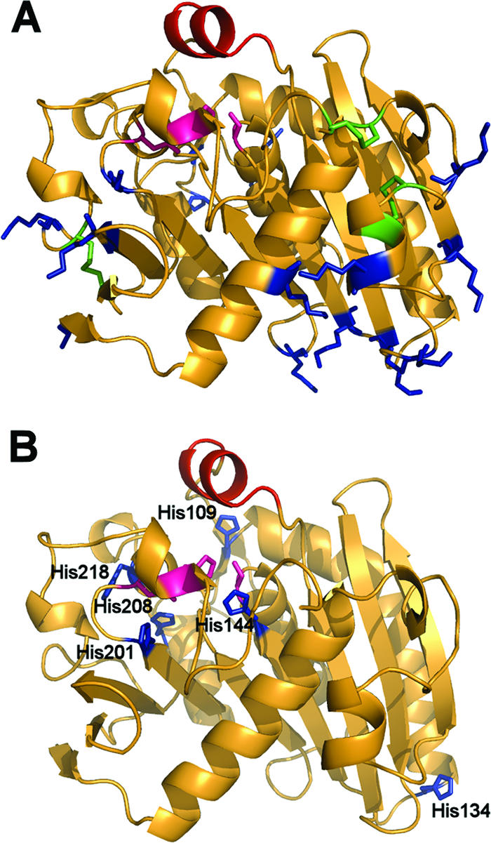
FIG. 1.

(A) Visualization of the target residues on the ROL. The lid (red), the active site (pink), the lysine groups (blue), and the cysteine groups forming disulfide bonds (green) are highlighted. (B) The lid (red), the active site (pink), and the histidine groups (blue) are highlighted.
