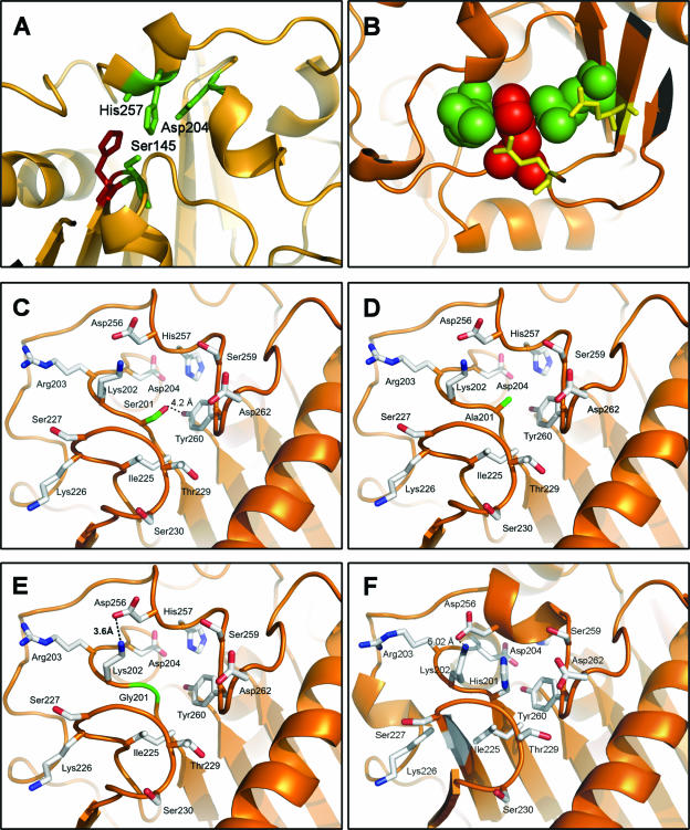
FIG. 6.
(A) His144 (in red) is adjacent to the catalytic serine. The catalytic triad Ser145, Asp204, and His257 is shown in green. (B) His218 (shown as red spheres) is “sandwiched” between Arg179 and Arg198 (shown as green spheres), which, in turn, establish salt bridge interactions with Glu222 and Glu240 (shown in yellow). (C to E) Local environment of H201X mutants. The ribbon diagrams show the ROL structure orange. The arrangement of the mutated His201 residues for Ser, Ala, or Gly (shown in green) is presented with the remaining residues defining the region (shown in light gray). (F) Wild-type ROL.

