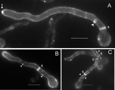
FIG. 3.
PalI localizes to the plasma membrane. Epifluorescence microscopy using a filter set specific for GFP of germlings cultured in acidic WMM. (A) With ethanol as the sole carbon source. The arrow indicates strong labeling in the position corresponding to the Spitzenkörper. (B and C) Germlings germinated in 0.02% glucose and shifted to WMM with 1% ethanol for 3 h. Membrane-associated punctate structures are indicated by arrowheads, whereas septae are indicated by s. Bars, 5 μm.

