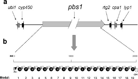
FIG. 1.
pbs1 locus of H. atroviridis and modular organization of the predicted PBS1 protein. (a) Chromosomal arrangement of the pbsl locus: ubi1, UbiA-like prenyltransferase; cyp450, cytochrome P450 subfamily protein; pbs1l, 19-mer peptaibol synthetase; rgt2, Rtg2-like protein; cpa1, Ca2+/H+ antiporter; lyp1, lysine permease. With the exception of pbs1, gene lengths are drawn to scale. (b) Modules within the PBS1 protein, numbered consecutively from 1 to 19. The domains within the modules are indicated as follows: gray cylinder, ketoacyl synthetase; black triangle, acyl transferase; white square, condensation domain; black circles: adenylation domains; gray triangles, thiolation domain; white arrow, alcohol dehydrogenase. The amino acids activated by the individual adenylation domains are indicated in the one-letter code (U, α-aminoisobutyric acid; J, isovaline).

