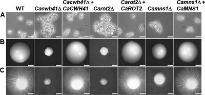
FIG. 1.
Cell and colony morphology in the Cacwh41Δ, Carot2Δ, and Camns1Δ null mutants. (A) Cell morphology after growth at 30°C for 16 h in YPD medium, demonstrating clumping of cells in the Cacwh41Δ (HMY19), Carot2Δ (HMY12), and Camns1Δ (HMY5) null mutants. Scale bars, 10 μm. (B and C) Colony morphology after 5 days growth at 30°C on YPD agar plates (B) or solid Spider medium (C). Scale bars, 1 mm.

