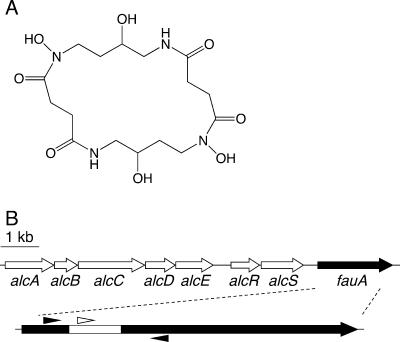
FIG. 1.
Molecular structure of alcaligin and the spatial organization of the Bordetella alcaligin siderophore system gene cluster. (A) Molecular structure of alcaligin. (B) Genetic map of the alcaligin system gene cluster. Arrows indicate the transcriptional orientations and spatial limits of genes. An arrow representing the 2.2-kb fauA coding sequence is enlarged below, and the DNA region deleted in the construction of the nonpolar ΔfauA2 mutant strain PM11 is indicated by the white segment. The small arrowheads flanking the fauA region deleted in PM11 approximate the positions of oligonucleotide primers used in cPCR to determine strain ratios in bacterial populations recovered from infected mice. The small white arrowhead above the deleted region represents the position of the oligonucleotide probe used in confirmatory colony hybridizations to distinguish fauA+ bacteria from ΔfauA2 mutants in the infection output. A 1-kb scale bar is shown.

