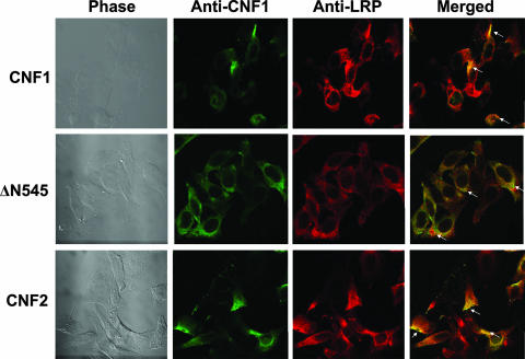
FIG. 6.
Colocalization of CNF1 and LRP on HEp-2 cells. CNF1, ΔN545, and CNF2 were bound to HEp-2 cells, and cells were subsequently stained with polyclonal anti-CNF1 sera, anti-LRP sera, and the corresponding fluorescently labeled secondary antibodies to allow visualization of toxin (green; Alexa 488) and LRP (red; Alexa 555). Confocal microscopy was used to demonstrate colocalization of the toxin and LRP receptor, as indicated by a yellow color (arrows) in the merged panels. Images were taken at a magnification of ×100.

