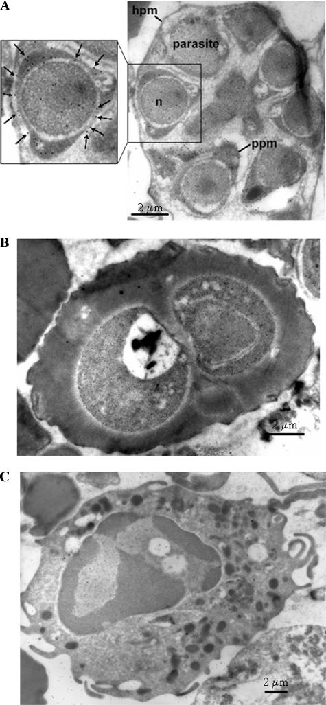
FIG. 5.
IEM image using mouse anti-r-Pfen antiserum at a 1:100 dilution with a P. falciparum-infected red cell at segmented schizont stage (A), a P. falciparum-infected cell at young trophozoite stage (B), and a human leukocyte (C). In panel A, the presence of enolase on the parasite plasma membrane (ppm) is marked with arrows. Hpm, host cell plasma membrane; n, nucleus.

