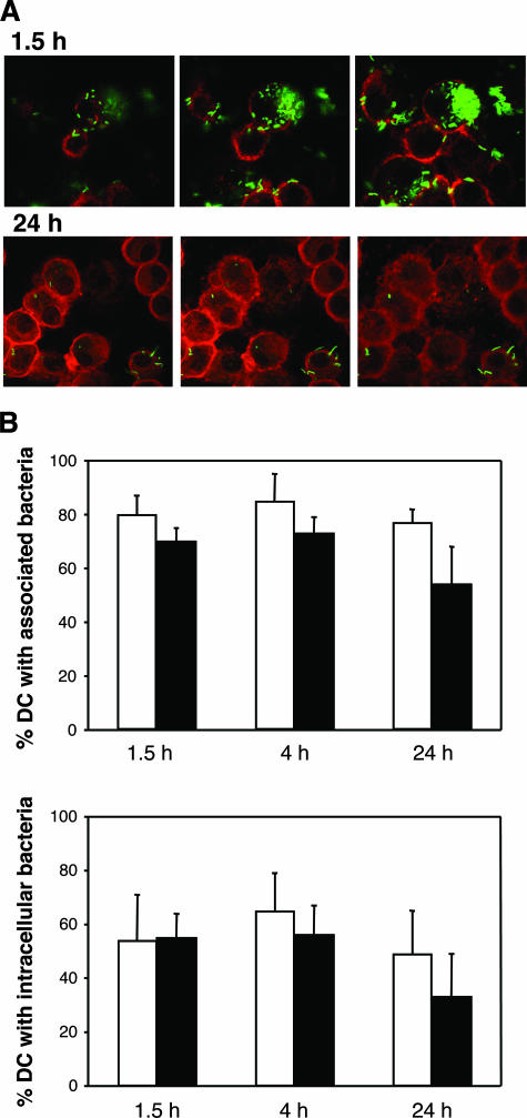
FIG. 4.
Confocal microscopy of DC infected with GFP-expressing H. ducreyi. (A) Optical sectioning of DC that had been incubated with H. ducreyi for 90 min and 24 h. (B) Percentages of DC that had associated (top) or internalized (bottom) bacteria under opsonized (white bars) and nonopsonized (black bars) conditions. The data represent the means ± SDs for assays performed with DC from three donors.

