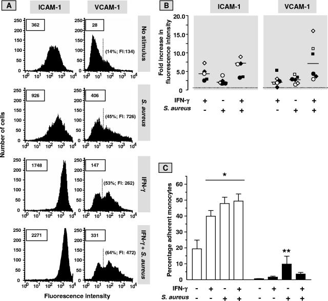
FIG. 6.
IFN-γ does not avert the S. aureus-induced endothelial inflammatory response. Monolayers of ECs, cultured for 72 h in culture medium alone or supplemented with 500 U/ml IFN-γ were exposed to S. aureus (EC to bacteria ratio, 1:100) or incubated with plain medium for 1 h, washed, treated with lysostaphin, and cultured for an additional 23 h in culture medium without antibiotics. Then, the cells were prepared for analysis by fluorescence-activated cell sorter (A and B) or used in the monocyte adhesion experiments (C). (A) A typical analysis by flow cytometry of endothelial CD54 (ICAM-1) and CD106 (VCAM-1) surface expression. Boxed numbers represent the mean fluorescence intensities expressed in arbitrary units of all analyzed ECs. Mean background fluorescence of cells incubated with the phycoerythrin-conjugated MAb ranged only from 15 to 30. The proportions of the cells that expressed VCAM-1 above the background level and their mean fluorescence intensities (FI) are depicted in parentheses. (B) Levels of expression of ICAM-1 and VCAM-1 in five and six separate experiments, respectively, with ECs from different donors were calculated relative to the median level of expression (set at level 1; dotted line) on untreated cells. Identical symbols represent data from the same experiment. Horizontal bars represent the medians of the separate experiments. (C) Monocytes were prepared and preincubated with plain medium (open bars) or with a panel of MAb directed against the β1- and β2-integrin molecules (closed bars) to block their interaction with endothelial VCAM-1 and ICAM-1, respectively, as described in Materials and Methods. Monocytes were allowed to adhere to the prepared EC monolayers for 30 min, and the percentage of EC-bound monocytes was determined thereafter. Data are given as means ± standard deviations for three separate experiments with cells from different donors. A paired t test was used to evaluate the data. *, P < 0.0001 relative to control ECs not treated with IFN-γ, bacteria, and MAb; **, P < 0.05 relative to one of the other EC cultures with MAb present.

