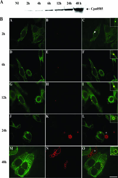
FIG. 5.
Cpn0585: kinetics of expression and colocalization with Rab11A in C. pneumoniae-infected cells. (A) Expression of Cpn0585 at various time points after C. pneumoniae infection. A total of 40 μg of protein from C. pneumoniae-infected HEp-2 cell detergent-solubilized extracts prepared at each of the indicated time points after C. pneumoniae infection was separated by SDS-PAGE and then transferred to a nitrocellulose membrane. Immunoblotting with rabbit anti-Cpn0585 antibodies detected a protein of an estimated molecular mass of 77 kDa as early as 2 h postinfection. Extracts from uninfected HEp-2 cells (NI) were used as a negative control. (B) HEp-2 cells transiently expressing EGFP-Rab11AQ70L were infected with C. pneumoniae at an MOI of 20, and at the indicated time points after infection the cells were fixed, stained with anti-Cpn0585 antibodies, and analyzed by laser scanning confocal microscopy. The signals from EGFP-Rab11AQ70L (green; A, D, G, J, and M) and anti-Cpn0585 stain (red; B, E, H, K, and N) were merged (C, F, I, L, and O). In the insets of panels with the merged images, the colocalization of Cpn0585 and EGFP-Rab11AQ70L is clearly noted as early as 2 h postinfection (arrow in panel C). Asterisks indicate C. pneumoniae-infected cells without EGFP-Rab11AQ70L expression. Bar, 10 μm. The data are representative of three experiments.

