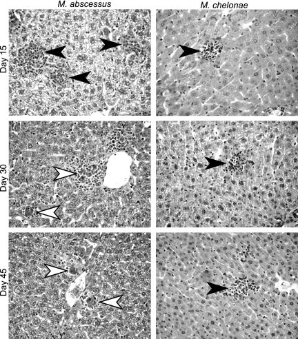
FIG. 6.
Histopathological analysis of liver sections of infected Ifngr1−/− mice. Animals were inoculated by the intravenous route with 107 CFU of M. abscessus or M. chelonae. HES-stained liver sections (magnification, ×400) obtained on days 15, 30, and 45 after M. abscessus or M. chelonae challenge are shown. The filled arrowheads indicate undifferentiated inflammatory infiltrates; the open arrowheads indicate differentiated granulomas featuring an epithelioid core and lymphocytic cuff. Slides were obtained from one of three independent experiments.

