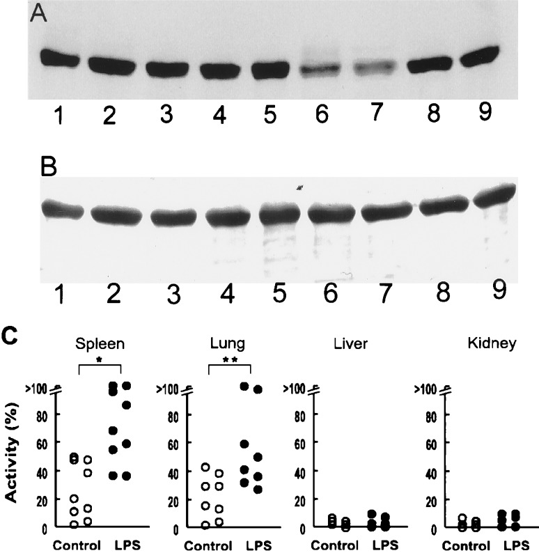
Figure 1.
Effects of LPS treatment on the activity of tissue homogenates to decrease nitrotyrosine content of nitrated BSA. (A) A representative Western immunoblot of nitrated BSA (1 mg/ml) that was incubated at 37°C for 10 min with tissue homogenates (2.5 mg/ml) of spleen (lanes 1, 6), lung (lanes 2, 7), liver (lanes 3, 8), and kidney (lanes 4, 9) from rats treated with LPS (20 mg/kg) for 24 h (lanes 6–9) or vehicle (control, lanes 1–4). Nitrated BSA was also incubated without tissue homogenate (lane 5). (B) Coomassie brilliant blue staining of a transferred membrane. Lanes are the same as in A. (C) Denitrating activity of tissues from (6–9) control rats (open symbols) and rats treated with LPS (solid symbols). Each symbol represents a different animal. Values are plotted as percent of maximal activity to decrease nitrated BSA. ∗, P < 0.01; ∗∗, P < 0.05, compared with control animals.

