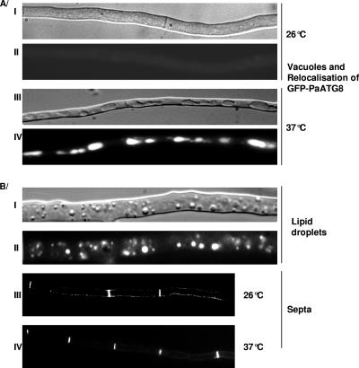
FIG. 4.
Cytological effects of HET domain expression. All experiments were conducted as described elsewhere (37). (A) Induction of autophagy by HET domain expression. A WT strain expressing a GFP-PaATG8 fusion protein and the HET domain was grown on complete medium at 26°C for 24 h and transferred to 37°C for 4 h to induce expression of the HET domain. (I and III) light microscopy using a Nomarsky filter; (II and IV) fluorescence microscopy. (B) Production of lipid droplets and septa by HET domain expression. A HET domain-expressing transformant was grown for 24 h on complete medium at 26°C and transferred for 2 h, 4 h, or 6 h to 37°C before observation. After 2 h of incubation at 37°C, lipid droplets were visible. Pictures were taken after 6 h of incubation at 37°C. (I and III) Light microscopy with a Nomarsky filter; (II) Nile Red staining of lipid droplets; (IV) Congo Red staining of septa observed under fluorescence microscopy.

