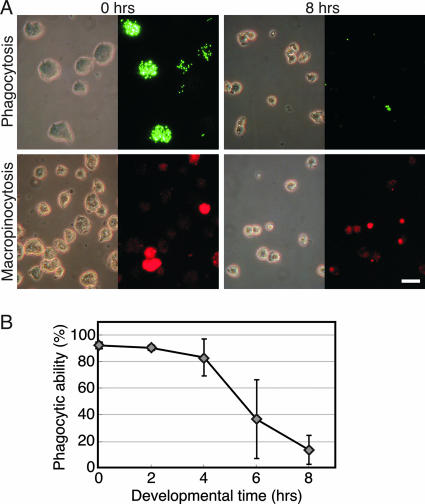
FIG. 2.
Phagocytosis and macropinocytosis are decreased during development. (A) Cells were harvested from growth media at 0 h or after 8 h of development on KK2 agar as indicated, washed, and incubated with fluorescent Dragon Green-conjugated polystyrene beads for phagocytosis or Texas Red dextran for macropinocytosis assays, as indicated on the left. Photographs were taken of representative fields by use of phase-contrast microscopy (left) and fluorescence microscopy (right). Scale bar, 20 μm. (B) Phagocytosis was quantified, and data are presented as averages ± SD of four independent replications.

