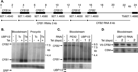
FIG. 5.
(A) Map of the region of chromosome 1 containing the CFB genes. Black blocks represent the coding regions, indicated by gene names above and locus numbers below. The scale above is in kilobases. The predicted mRNAs are indicated below the open reading frames, the spliced leader being indicated by a black dot. (B) Regulation of CFB mRNAs after overexpression of TbUBP2 or expression of TcUBP1. Northern blots of bloodstream or procyclic trypanosomes with tetracycline-inducible TcUBP1 or TbUBP2 were hybridized with DNA derived from positive array spots (see Table 1). Signal recognition particle (SRP) RNA served as a loading control. Cells with overexpression (+) were incubated with tetracycline for 24 h. (C) Effects of RNA interference downregulation of either PEX2 or TbUBP1/2. The cultured cells containing tetracycline-inducible constructs were incubated for either 24 h (1 day) or 48 h (2 days) with tetracycline. Note that after 2 days of PEX2 RNAi, the cells were dying and mRNA was disappearing. (D) Effect of TbUBP1/2 RNAi on the level of V5-tagged CFB2 in bloodstream cells. The loading control is a cytosolic marker protein. The results from two independent V5-tag clones are shown.

