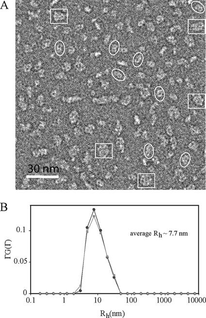
FIG. 2.
Analysis of wild-type untagged HMW1B by electron microscopy and dynamic light scattering suggests a dimer in DDM solution. (A) An electron micrograph of HMW1B particles negatively stained with phosphotungstic acid. Electron microscopy was performed with a Jeol-1200EX microscope, and images were recorded on a Gatan 791 charge-coupled device camera. White ellipses mark examples of dimers, and white rectangles mark examples of dimers of dimers. (B) Size distribution of HWM1B in detergent micelles as derived from the CONTIN analysis of the dynamic light scattering measurement. The two curves represent two separate measurements.

