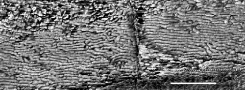
FIG. 2.
AFM image of Oscillatoria sp. strain A2. The scan was performed on a sample that had been transferred from an agar plate to a glass slide and was, consequently, dehydrated when scanned. Parts of two cells are visible, with a cell septum running vertically down the center of the micrograph. Scale bar, 500 nm.

