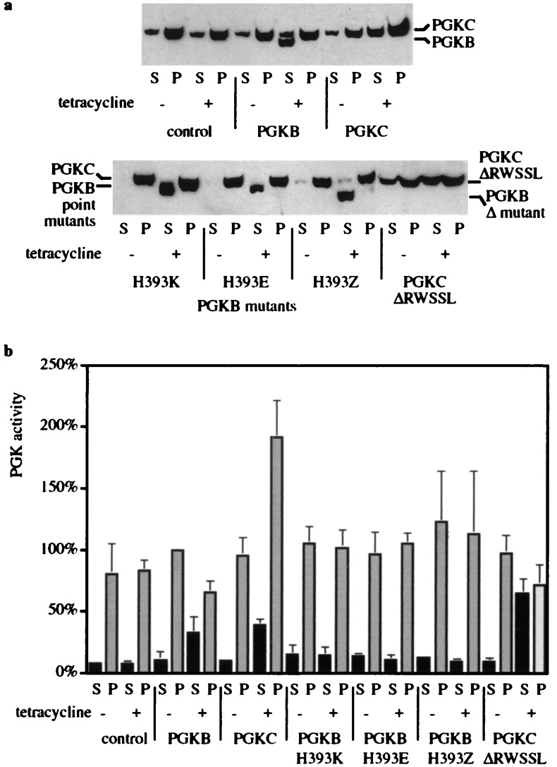
Figure 3.
Location (a) and activity (b) of various versions of PGK inducibly expressed in bloodstream forms. Cultures (30 ml) initiated at 5 × 10⁵ cells/ml in the presence or absence of 5 μg/ml tetracycline were grown for 24 h; aliquots of 107 cells were then subjected to cell fractionation with digitonin. S, supernatant; P, pellet. (a) Western blot of TCA-precipitated protein using anti-PGK antibody. (b) Enzyme activities were measured from fractions equivalent to 4 × 10⁶ cells. Results were calculated relative to the pellet fraction of the PGKB cells without tetracycline, set to 100%, and are the mean ± standard deviation from three or four independent experiments. Cell lines contained the PGK coding regions followed by the VSG 3′ UTR.

