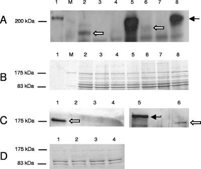
FIG. 4.
Expression analysis of HrpA proteins by Western immunoblot assays. Proteins were separated on a 7.5% polyacrylamide gel, transferred to a nitrocellulose membrane, and probed with polyclonal antibody RαNMB1779. The arrows indicate the processed (white arrows) and unprocessed (black arrow) HrpA proteins. (A) Western blot of whole-cell lysates of strain MC58, strain 2517, and their respective ΔhrpA and ΔhrpB mutants. Lanes: 1, purified HrpA; M, molecular mass standard (in kilodaltons); 2, MC58 (wild type); 3, MC58 ΔsiaD ΔhrpA; 4, MC58 ΔgalE ΔhrpA; 5, MC58 ΔsiaD ΔlgtA ΔhrpB; 6, 2517; 7, 2517 ΔhrpA; 8, 2517 ΔhrpB. In strain MC58, ΔhrpA denotes simultaneous deletion of NMB0497 and NMB1779, whereas ΔhrpB denotes simultaneous deletion of NMB0496 and NMB1780. (B) Coomassie blue-stained gel of whole-cell lysates used in panel A. (C) Western blot of culture supernatants of strain 2517 and its respective ΔhrpA and ΔhrpB deletion mutants as well as the double deletion mutant ΔhrpA ΔhrpB in comparison to whole-cell lysate of strain 2517 ΔhrpB. Lanes: 1, 2517; 2, 2517 ΔhrpA; 3, 2517 ΔhrpB; 4, 2517 ΔhrpA ΔhrpB; 5, whole-cell lysate of strain 2517 ΔhrpB; 6, 2517. (D) Coomassie blue-stained gel of culture supernatants used in panel C.

